
Hip Dysplasia in Women: The Diagnosis Many Don’t Receive Until Adulthood
If you have lived with persistent hip or groin pain for years—and have been told that your hip is “tight,” your hip flexors are weak, your ITB is the problem, or that you simply need to stretch more or strengthen the gluts – there may be another explanation worth considering.
Hip dysplasia can go undiagnosed until adulthood.
For some women, symptoms do not become significant until their teens, 20s, 30s, or even later. Others have spent years treating hip, back, SI joint, or pelvic pain without anyone stepping back to ask an important question:
Is the shape and stability of the hip joint contributing to the problem?
Hip dysplasia is not something you can diagnose from symptoms alone. But recognizing its characteristic presentation can help you know when a more thorough hip evaluation may be warranted.
What Is Hip Dysplasia?
©2026. Ginger Garner. All rights reserved.
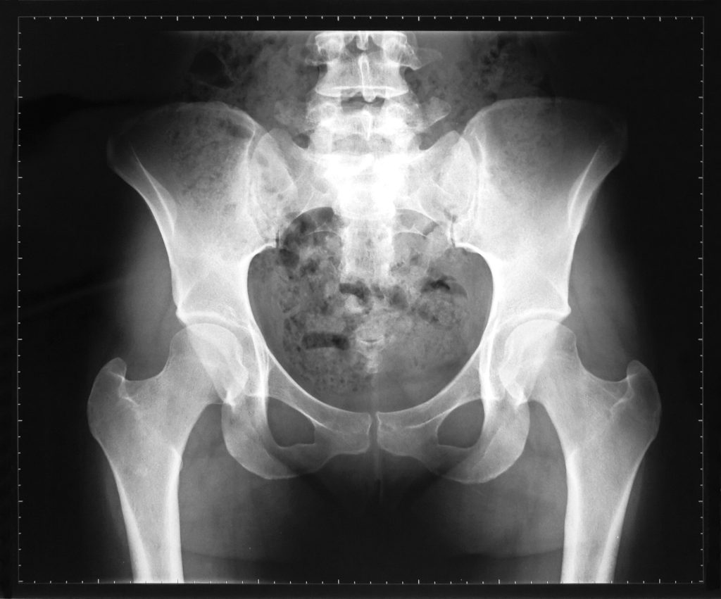
The hip is a ball-and-socket joint. The “ball” is the head of the femur, or thigh bone. The “socket” is the acetabulum, which is part of the pelvis. In a well-developed hip, the socket provides adequate coverage and support for the head of the femur. In acetabular or developmental hip dysplasia, the socket may be too shallow or developed in a way that its position provides insufficient coverage of the femoral head.
That changes how forces travel through the joint. Instead of having the bony architecture provide optimal stability, the labrum, joint capsule, muscles, tendons, and surrounding soft tissues may be asked to do more work to stabilize the hip.
Over time, that can contribute to pain, labral injury, instability, and eventually osteoarthritis in some patients.
Hip dysplasia exists along a spectrum. A hip does not have to be dislocated—or look dramatically abnormal—to cause symptoms.
Why Can Hip Dysplasia Be Missed Until Adulthood?
Developmental dysplasia of the hip is associated with childhood, but not every dysplastic hip is identified during childhood screening. Infant screening is particularly focused on clinically unstable hips and children with recognized risk factors such as breech presentation and family history. The American Academy of Orthopaedic Surgeons notes that even childhood screening and imaging strategies have limitations.
Some people therefore reach adulthood with a hip that is stable enough to function but structurally undercovered. Symptoms may emerge as activity demands increase or as the tissues surrounding the joint become less able to compensate.
And women deserve particular attention. A 2024 systematic review examining radiographic hip dysplasia in asymptomatic adults found dysplasia in approximately 3.8% of females compared with 2.7% of males in studies reporting prevalence by sex.
The symptoms can also be surprisingly nonspecific. Research asking adults with acetabular dysplasia to describe their experiences identified patterns involving pain, gait and posture, childhood or family hip history, and unusual hip-joint sensations. In other words, hip dysplasia does not always announce itself with an obvious “hip problem.”
What Does Hip Dysplasia Feel Like?
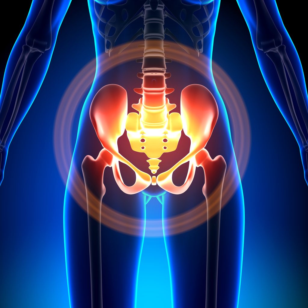
Symptoms vary, but common complaints can include:
©2026. Ginger Garner. All rights reserved.
- Deep groin or anterior hip pain
- Lateral hip pain
- Buttock pain
- Pain with prolonged standing or walking
- Pain after exercise or running
- Pain with squatting or deeper hip flexion
- Clicking, catching, popping, or grinding
- A feeling that the hip is unstable or needs to “shift”
- Fatigue or aching around the hip
- Difficulty standing on one leg
- Low-back or pelvic pain accompanying the hip symptoms
Recent international consensus work on hip instability identified anterior hip pain with daily activities, mechanical symptoms such as popping or clicking, and subjective feelings of looseness or instability among important clinical features.
Female sex and hip dysplasia were also recognized as risk factors for atraumatic hip instability.
None of those symptoms automatically means you have dysplasia. They do mean that persistent hip symptoms deserve a careful evaluation rather than simply being treated indefinitely as “tight muscles.”
The Hip Dysplasia–Pelvic Floor Connection
This is one of the most overlooked parts of the conversation.
Your pelvic floor does not function independently of your hips.
The deep hip muscles, pelvic floor, abdominal wall, diaphragm, pelvis, and spine work together to help control load and movement through the lumbopelvic region. When the bony structure of the hip provides less stability, the surrounding muscles may increase their activity in an effort to create stability.
That compensation can sometimes be useful. But over time, a strategy of constantly “holding” around the hip and pelvis may also contribute to pain, guarding, altered movement, and difficulty relaxing the pelvic floor.
Research supports a broader relationship between lumbopelvic pain and pelvic-floor dysfunction, although we do not yet have evidence showing that hip dysplasia itself directly causes pelvic-floor dysfunction in every patient.
Women with chronic pelvic pain have also been shown to demonstrate higher rates of pelvic-floor tenderness and impaired pelvic-floor relaxation than women without pelvic pain.
Clinically, that distinction matters. If you have both hip and pelvic symptoms, treating only the pelvic floor—or only the hip—may miss part of the picture. For some patients I see, the question becomes:
Is the pelvic floor actually the primary problem, or is it working overtime because the hip and pelvis need more support?
This is especially important when pelvic pain coexists with groin pain, painful intercourse, deep pelvic tension, buttock pain, hip instability, or difficulty relaxing the pelvic floor.

©2021. Pelvic Guru, LLC. Used with kind permission.
How Is Hip Dysplasia Diagnosed?
Diagnosis typically involves a combination of:
Your history. When did the symptoms begin? What aggravates them? Were you breech? Is there a family history of hip problems or early hip replacements? Have you always felt unusually flexible or unstable?
Physical examination. Hip range of motion, strength, movement control, gait, functional movement, and signs of instability can all provide useful information.
Imaging. X-rays are generally essential for evaluating the bony structure of the hip. Measurements of acetabular coverage and orientation can help determine whether dysplasia is present. A common measurement used, but certainly not the only one, is the Lateral Central Edge Angle (LCEA).
MRI may also be appropriate when labral or cartilage injury is suspected. However, it isn’t 100% accurate and can have a false positive (it says you have a tear when you don’t) or negative (it says you don’t have a tear when you do). Interpretation is also highly dependent on who is looking at the MRI.
So it’s especially important that you see a hip dysplasia and/or hip preservation specialist for proper diagnosis. Importantly, an MRI showing a labral tear does not necessarily tell us why the labrum became injured or if it is even important or causing the primary pain.
Sometimes the bigger question is what is happening structurally underneath it.

©2026. Ginger Garner. All rights reserved.
Can Physical Therapy Help Hip Dysplasia?
Yes—but rehabilitation has to respect the structure of the hip. Physical therapy cannot make a shallow acetabulum deeper. What we can do is improve how effectively your body manages the forces going through that joint. Rehabilitation may include:
- Strengthening the muscles that support the hip and pelvis (see the gluteal sling figure above)
- Improving movement and load management
- Addressing gait and functional mechanics
- Building trunk and lumbopelvic control
- Reducing unnecessary muscular guarding
- Modifying activities that repeatedly provoke symptoms
- Treating coexisting pelvic-floor dysfunction when present
- Helping patients understand which movements their hip tolerates well—and which it does not
- If fascial limitations are inhibiting muscle strength, timing, and control
Importantly, treatment should not automatically focus on gaining more hip mobility. And, physical therapy should not only address strength. In my practice I often find myofascial restriction to be a primary pain generator. If your problem involves instability or inadequate structural coverage, aggressively stretching can on an already mobile hip is not always the answer. See the video below regarding hip flexor stretching.
Sometimes the goal isn’t more mobility. It’s better control and better load management.
What About Surgery?
Not every person with hip dysplasia needs surgery. Treatment depends on the degree of dysplasia, symptoms, activity demands, joint cartilage, age, associated labral or cartilage injury, and response to conservative management. When symptomatic dysplasia is significant and the joint is otherwise appropriate for preservation, a procedure called a periacetabular osteotomy (PAO) may be considered. PAO changes the orientation of the acetabulum to improve coverage of the femoral head.
Research examining adults undergoing PAO shows meaningful improvements in pain, function, and quality of life following surgery, although recovery is substantial and appropriate patient selection is important. For this reason, someone with suspected dysplasia may benefit from evaluation by an orthopedic surgeon who specializes specifically in hip preservation, rather than simply general hip surgery.
When Should You Ask About Hip Dysplasia?
Consider asking for a more comprehensive hip evaluation if you have persistent hip or groin pain—particularly if you are a younger or middle-aged woman—and:
- Treatment repeatedly helps temporarily but the pain returns
- You have deep groin pain or mechanical clicking or catching
- Your hip feels unstable rather than simply stiff
- You have always been extremely flexible
- Hip pain accompanies pelvic-floor or pelvic pain, and/or painful intercourse
- Symptoms increase with walking, running, standing, squatting, or single-leg activities
- You have been diagnosed with a labral tear without a clear explanation for why it developed
- You have been told your imaging is “normal,” but your symptoms continue
There are many causes of hip and pelvic pain, and hip dysplasia is only one of them. But it is an important diagnosis not to miss. When a structural hip problem is driving symptoms, repeatedly treating the painful tissues without recognizing the underlying mechanics may never fully solve the problem.
And for women who have spent years trying to understand unexplained hip and pelvic pain, simply asking the right question can sometimes change the entire direction of care: Could the hip joint itself be part of the reason I hurt?
What Do Hypermobility and Ehlers-Danlos Syndrome Have to Do With Hip Dysplasia?
There is another group of patients in whom I pay particularly close attention to hip stability: people with generalized joint hypermobility, hypermobility spectrum disorder (HSD), or hypermobile Ehlers-Danlos syndrome (hEDS).
These conditions are not the same thing as hip dysplasia, and having hypermobility does not mean you have a dysplastic hip.
But they can overlap.
Hip stability depends on more than the shape of the bones. It also depends on the joint capsule, ligaments, labrum, and muscles surrounding the hip. Hip dysplasia can decrease the amount of bony coverage supporting the femoral head. Generalized connective-tissue laxity can create an additional challenge by allowing more movement through the joint capsule and supporting tissues.
That means some patients may have both less bony stability and more soft-tissue laxity. Research on hip microinstability recognizes both acetabular dysplasia and generalized connective-tissue disorders such as Ehlers-Danlos syndrome as potential contributors to excessive or poorly controlled motion at the hip joint.[1–4]
This combination can be especially important in women who have spent years being described as simply “very flexible.” Flexibility is not always the same thing as functional mobility.
A person may have tremendous range of motion and still lack the ability to control the femoral head effectively within the socket. In that situation, stretching an already hypermobile hip may actually be counterproductive.
Symptoms may include:
- Deep groin or anterior hip pain
- Clicking, popping, or catching
- A sensation that the hip shifts or does not feel secure
- Pain after prolonged walking, standing, or exercise
- Symptoms with positions requiring large ranges of hip motion
- Muscle fatigue or chronic tightness around the hips and pelvis
- Pelvic, buttock, or low-back pain
- Repeated injuries despite being “flexible”
Studies of hip microinstability have found that the majority of reported patients are women, and generalized ligamentous laxity is recognized as an important factor when evaluating an unstable or painful hip.[2,3] For patients with HSD or hEDS, this distinction is particularly important:
The problem may not be that the hip needs more mobility. The problem may be that the hip needs better stability, fascial glide, muscular support, more complex motor patterns, bony alignment, proprioception, and load tolerance. That can fundamentally change the rehabilitation plan.
The Hip Dysplasia–Pelvic Floor Connection
This is one of the most overlooked parts of the conversation. Your pelvic floor does not function independently of your hips. The deep hip muscles, pelvic floor, abdominal wall, diaphragm, pelvis, and spine work together to manage pressure, movement, and load through the lumbopelvic region.
When the bony structure of the hip provides less stability—or when connective-tissue laxity allows excessive joint motion—the surrounding muscles may increase their activity in an effort to create stability.
That compensation may be helpful initially. But continually using muscle tension as a stability strategy can also contribute to fatigue, guarding, pain, and difficulty relaxing the pelvic floor. This becomes especially relevant for patients with hypermobility and EDS.
Women with EDS report high rates of pelvic-floor symptoms. In a large international survey of 1,303 women with EDS, 71% reported pelvic pain, while urinary symptoms, prolapse symptoms, and dyspareunia were also common.[5] A 2026 study examining women with hEDS or hypermobility spectrum disorder presenting with genito-pelvic pain also found a substantial overlap between hypermobile features, vulvovaginal pain, sexual dysfunction, and pelvic-floor-related pain presentations.[6]
Those studies do not prove that hip dysplasia or hypermobility directly causes pelvic-floor dysfunction. But they reinforce something clinically important:
Hip stability, connective-tissue function, and pelvic-floor symptoms should not always be evaluated as separate problems. For someone with hip dysplasia plus hypermobility, the pelvic floor may sometimes become part of a broader stabilization strategy. That is why simply telling a patient to “relax the pelvic floor” may not be enough. Sometimes we first need to ask: Why does the nervous system believe it needs that much muscular tension to feel stable? And in some patients, the answer includes the hip.
Rehabilitation Is Different When Hypermobility Is Part of the Picture
Physical therapy for hip dysplasia should already emphasize control and load management rather than simply increasing range of motion. When generalized hypermobility, HSD, or hEDS is also present, that principle becomes even more important. Rehabilitation may emphasize:
- Progressive hip and trunk strengthening
- Muscular endurance rather than maximal flexibility
- Proprioception and joint-position awareness
- Control through mid-range rather than repeatedly moving into end-range
- Gradual exposure to functional loading
- Gait and movement strategies
- Pelvic-floor coordination when pelvic symptoms are present
- Reducing unnecessary muscular guarding without removing the stability the patient needs
- Avoiding aggressive stretching when the joint already has excessive mobility
This is one reason a generic stretching program may fail a hypermobile patient. A muscle that constantly feels “tight” may not actually be short. It may be working overtime to stabilize a joint that does not feel secure. The goal is not simply to make that muscle relax. The goal is to give the entire system a better strategy for stability.
Looking for help? First consults are always free at www.garnerpelvichealth.com
Select References
- Guimarães JB, et al. Hip Microinstability: New Concepts and Comprehensive Imaging Evaluation. Radiographics. 2025;45(7):e240134. doi:10.1148/rg.240134.
- Cohen D, et al. Hip microinstability diagnosis and management: a systematic review. Knee Surg Sports Traumatol Arthrosc. 2023.
- Mei-Dan O, et al. Microinstability of the Hip and the Splits Radiograph. Orthopedics. 2016.
- Hip Microinstability: Current Concepts in Diagnosis, Surgical Management, and Outcomes—A Narrative Review. 2025.
- Kciuk O, Li Q, Huszti E, McDermott CD. Pelvic floor symptoms in cisgender women with Ehlers-Danlos syndrome: an international survey study. Int Urogynecol J. 2022.
- Barton L, et al. Characterizing sexual dysfunction in females with hypermobile Ehlers Danlos syndrome or hypermobility spectrum disorder and genito-pelvic pain through cross-sectional analysis. Rheumatol Int. 2026;46(7):145. doi:10.1007/s00296-026-06161-w.


